Dental imaging · Hoppers Crossing

Seeing what an examination cannot show.

Digital X-rays, panoramic OPG and 3D CBCT imaging at our Sayers Road practice, used only when the information will help diagnose or plan your care.

  • Intraoral X-rays
  • Panoramic OPG
  • 3D CBCT
Digital dental X-ray
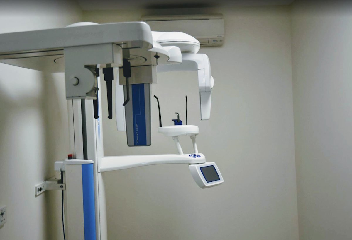
Dental imaging room at Sayers Dental
Imaging room · Sayers Dental
Why imaging is used

Some problems are hidden from view.

Decay between teeth, infection at the tip of a root, bone loss from gum disease and wisdom teeth still sitting in the jaw are often invisible during an examination. Imaging lets your dentist see these areas, so a diagnosis rests on evidence rather than guesswork.

Our Hoppers Crossing practice has digital intraoral X-rays and 3D OPG/CBCT imaging on site. Digital sensors generally need less radiation than older film, and images appear on screen within moments, so your dentist can show you exactly what they are looking at. Imaging can help identify:

  • Decay between teeth or under existing fillings
  • Infection or inflammation around a root
  • Bone levels around teeth affected by gum disease
  • The position of wisdom teeth and developing teeth
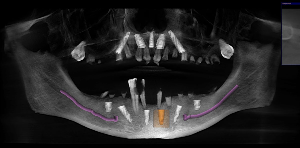
  • Bone volume and nerve position before implant treatment

Images are always read alongside your examination and health history before anything is recommended.

The Sayers Dental team
Hoppers Crossing
• S •
Types of imaging

Three kinds of image, each with a purpose.

The type of image depends on the question your dentist needs answered. Many visits need none at all.

01.Intraoral

Bitewing and periapical X-rays

Small images taken with a sensor placed inside the mouth. Bitewings show the upper and lower back teeth together and are used to check for decay between them. Periapical images show one or two teeth from crown to root tip.

02.Panoramic

OPG X-ray

An orthopantomogram (OPG) captures all the teeth, both jaws and the jaw joints in one wide image. The machine moves around your head, and the exposure takes around 20 seconds.

03.3D

CBCT scan

Cone beam computed tomography (CBCT) produces a three-dimensional view of the teeth, bone and nerve canals. It is mainly used to plan implants, complex extractions and some root canal treatment.

Your dentist will explain why an image is recommended, and what it is expected to show, before it is taken.

Radiation, in plain terms

Low doses, used only when justified.

Everyone receives a small amount of natural background radiation every day, from the ground, the air and even food. Dental X-rays add a little to this. As a general guide, a single intraoral X-ray is roughly similar to a day or so of natural background radiation, and a panoramic OPG to a few days.

A 3D CBCT scan involves a higher dose than a standard dental X-ray, and the dose varies with the size of the area scanned. That is why it is recommended only when the extra detail is likely to change how treatment is planned, and why the scanned area is kept as small as practical.

Dose comparisons are general estimates and vary with the equipment and the area imaged.

How we keep exposure low
Clinical need first
Images are taken only when the information is expected to benefit your care.
Digital sensors
Digital imaging generally needs less radiation than traditional film.
The smallest useful area
Scans are limited to the region your dentist needs to see.
Using existing images
Recent X-rays from another practice can sometimes be used instead of new ones, so bring them if you have them.
Pregnancy
Tell us if you are or may be pregnant, so your dentist can consider whether imaging can wait.
When imaging may be recommended

Common reasons for an image.

Your dentist decides on imaging case by case, based on your symptoms, history and any treatment being planned.
01.Check-ups

Routine monitoring

Bitewings at intervals based on your decay risk, to find problems between teeth early.

02.Pain

Toothache or swelling

To look for infection, a cracked root, or decay close to the nerve.

Having an image taken

Quick, simple and over in moments.

No special preparation is needed. Your dentist or dental assistant will guide you through each step.

Dental imaging room at Sayers Dental
  1. 01.

    Before

    Let us know if you are or may be pregnant, and remove glasses, earrings or other metal near your head and mouth if asked.

  2. 02.

    Positioning

    For intraoral X-rays a small sensor is placed in your mouth. For an OPG or CBCT you bite gently on a guide that keeps your head still.

  3. 03.

    The image

    You stay still for a few seconds while the image is taken. An OPG takes around 20 seconds as the arm moves around your head.

  4. 04.

    Your results

    Images appear on screen straight away, and your dentist talks you through what they show and what they mean for your care.

Common questions

Dental imaging questions.

General information about dental X-rays, OPGs and CBCT scans. Your dentist will advise whether any imaging is needed in your case.

Are dental X-rays safe?

Dental X-rays use low doses of radiation, and digital imaging generally needs less than older film. Any radiation carries a small risk, which is why images are taken only when the expected benefit to your care outweighs it. Please tell us beforehand if there is any chance you are pregnant.

What is an OPG?

An OPG (orthopantomogram) is a wide, single X-ray covering every tooth, both jaws and the jaw joints. It is often used to assess wisdom teeth, plan orthodontic or implant treatment, and look for problems in the jaw.

What is the difference between an OPG and a CBCT scan?

An OPG is a flat, two-dimensional image of the whole mouth. A CBCT scan is three-dimensional, showing the width and depth of bone and the position of nerves, which helps when planning implants or complex extractions. CBCT involves a higher dose, so it is used only when that extra detail is needed.

How often do I need dental X-rays?

There is no fixed schedule. Your dentist considers your age, your risk of decay and gum disease, any symptoms and any treatment being planned. Some people need bitewings every year or two, others less often.

Do I need to prepare for a dental X-ray?

No special preparation is needed. Remove glasses, earrings and other metal items near your head if asked, and tell us if you are or may be pregnant.

Does having a dental X-ray hurt?

No. Some people find the intraoral sensor a little awkward to bite on, especially towards the back of the mouth, but the image itself takes only a moment. Tell us if you have a strong gag reflex and we can adjust the approach.

Can I use X-rays from my previous dentist?

Often, yes. If you have recent images, ask your previous practice to send them to us before your visit. Your dentist will decide whether they are recent and clear enough to use.

The next step

Book an examination.

Imaging is arranged as part of an examination or treatment plan. Book a visit and your dentist will advise whether any images are needed.

Find us

Sayers Dental Aesthetics & Implants

1/483 Sayers Road, Hoppers Crossing VIC 3029

(03) 9749 1178 · admin@sayersdental.com.au

  • Tue to Thu9:00am to 5:00pm
  • Fri10:00am to 6:00pm
  • Sat9:00am to 4:00pm
  • Sun and MonClosed

Get directions →

Request a call back

Complete the form and our reception team will be in touch during business hours.

Your information is kept private and used only to respond to your enquiry. See our privacy policy. For urgent dental pain, please call us.